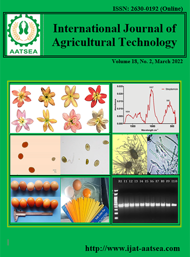
Antifungal and antioxidant activities of Ag@FeO-NPs@Chitosan preparation by endophyte Streptomyces aureofaciens
Main Article Content
Abstract
Biological control using beneficial microorganisms is nowdeveloped for reducing the plant pathogens. Nanoparticles with specific functions have been produced and represented an economical alternative to agochemical through the natural methods of nanoparticle formation. Biosynthesis of Ag@FeO-NPs@Chitosan preparation by Streptomyces aureofaciens was performed against some soil borne pathogenic fungi i.e. Fusarium oxysporum, F. solani, F. subglutinans, Rhizoctonia solani and air borne pathogenic fungi i.e. Botrytis cinerea, Colletotrichum gloeosporioides, Alternaria solani and Aspergillus niger . To conceive the formation of Ag@FeO-NPs@Chitosan, ultraviolet-visible (UV-Vis) spectroscopy for the biosynthesis of core–shell nanoparticle and fourier transform infrared (FTIR) spectroscopy analysis were done for constructing the structure of nano-composite. Scanning transmission electron microscopy (STEM), and scanning transmission electron microscopy (STEM) micrographs evidenced that the size of synthesized nanoparticles is less than 50 nm. As a result, the data showed that the produced core–shell hemtite @Ag nanoparticles are super-paramagnetic. The chitosan is formed in the outer shell of the formula spontaneously due to the presence of chitin in the fungus cell membrane. In vitro bioassay, Ag@FeO-NPs@Chitosan preparation by Streptomyces showed distinct antifungal against all plant pathogens, and inhibited the growth and spores germination. Antioxidant activities as reducing power measurement, DPPH (1,1-Diphenyl-2- picrylhydrazyl) radical scavenging activity, nitric oxide scavenging activity, ABTS free radical scavenging activity and total phenolics contents were greatly increased. Synthesized Ag@FeO-NPs@Chitosan preparation by S. aureofaciens is demonstrated remarkable potential for using as antifungal compound to combat plant diseases. Chitosan is a good source for the preparation of bionanoparticle materials for industry and applications
Article Details

This work is licensed under a Creative Commons Attribution-NonCommercial-NoDerivatives 4.0 International License.
References
Bautista-Baños, S., Hernández-López, M. and Barrera-Necha, L. L. (2000) Antifungal Screening of Plants of the State of Morelos, México against Four Fungal Postharvest Pathogens of Fruits and Vegetables. Mexican Journal of Phytopathology, 18:36-41.
Bhattacharyya, A., Duraisamy, P., Govindarajan, M., Buhroo, A. A. and Prasad, R. (2016.) “Nano-biofungicides: emerging trend in insect pest control,” in Advances and Applications through Fungal Nanobiotechnology, ed. R. Prasad (Cham: Springer International Publishing), 307-319.
Casillas, P. E. G., Pérez, C. A. M. and Gonzalez, C. A. R. (2012). Infra- red spectroscopy of functionalized magnetic nanoparticles, in Infrared Spectroscopy - Materials Science, Engineering and Technology, pp.405-420, INTECH Open Access Publisher.
Eid, M. M., El-Hallouty, S. M., El-Manawaty, M. and Abdelzaher, F. H. (2019.) Physicochemical Characterization and Biocompatibility of SPION@ Plasmonic@ Chitosan Core-Shell Nanocomposite Biosynthesized from Fungus Species. Journal of Nanomaterials, 2019. Retried from https://doi.org/10.1155/2019/4024958
Eid, M. M., El-Hallouty, S. M., El-Manawaty, M., Abdelzaher, F. H., Al-Hada, M. and Ismail, A. M. (2018). Preparation conditions effect on the physico-chemical properties of magnetic–plasmonic core–shell nanoparticles functionalized with chitosan: green route. Nano-Structures & Nano-Objects, 16, pp.215-223.
Farhat, M. G., Haggag, W. M., Thabet, M. S. and Mosa, A. A. (2018). Efficacy of Silicon and Titanium Nanoparticles Biosynthesis by some Antagonistic Fungi and Bacteria for Controlling Powdery Mildew Disease of Wheat Plants. International Journal of Agricultural Technology 14:661-674.
Fei Law , W., Leng Ser, H., Khan, Hong Chuah, L, Pusparajah, P., Gan Chan, K., Hing Goh, K. and Han Lee, L. (2017). The Potential of Streptomyces as Biocontrol Agents against the Rice Blast Fungus, Magnaporthe oryzae (Pyricularia oryzae). Frontiers in Microbiology, 17. Retried from https://www.frontiersin.org/articles/10.3389/fmicb.2017.00003/full
Folin, O. and Ciocalteu, V. (1927.) On tyrosine and tryptophane determinations in proteins. Journal of Biological Chemistry, 73:627-650.
Haggag, W. M. and Malaka, S. A. E. (2012). Application and Formulations of Streptomyces Aureofaciens and Pseudomonas Putida for Management of Grape Anthracnose and Grey Mould Diseases. European Journal of Scientific Research, 91:174-183.
Haggag, W. M. and Ali, R. R. (2019). Microorganisms for wheat improvement under biotic stress and dry climate. Agricultural Engineering International: CIGR Journal, 21: 118-126.
Hongtao, Cui., Yan, Liu. and Wanzhong, Ren. (2013). Structure switch between α-Fe2O3, γ-Fe2O3 and Fe3O4 during the large scale and low temperature sol–gel synthesis of nearly monodispersed iron oxide nanoparticles. Advanced Powder Technology, 24:93-97.
Kawahara, T., Izumikawa, M., Otoguro, M., Yamamura, H., Hayakawa, M. and Takagi, M, (2012). JBIR-94 and JBIR-125 antioxidative phenolic compounds from Streptomyces sp. R56-07. Journal of Natural Products, 75:107-110.
Kekuda, T. R. P., Shobha, K. S. and Onkarappa, R. (2010). Fascinating diversity and potent biological activities of Actinomycete metabolites. Journal of Pharmacy Research, 3:250-256.
Kumirska, J., Czerwicka, M., Kaczyński, Z., Bychowska, A., Brzozowski, K., Thöming, J. and Stepnowski, P. (2010). Application of spectroscopic methods for structural analysis of chitin and chitosan. Marine drugs, 8:1567-1636.
Lee, D. R., Lee, S. W., Choi, B. K., Cheng, J., Lee, Y. S., Yang, S. H. and Suh, J. W. (2014). Antioxidant activity and free radical scavenging activities of Streptomyces sp. strain MJM 10778 Asian Pacific Journal of Tropical Medicine, 7:962-967.
Liu, R. and Lal, R. (2015). Potentials of engineered nanoparticles as fertilizers for increasing agronomic productions. Science of the Total Environment, 514:131-139.
Marcocci, L., Maguire, J. J., Droy-Lefaix, M. T. and Packer, L. (1994). The nitric oxide-scavenging properties of Ginkgo biloba extract EGb 761. Biochemical and Biophysical Research Communications, 15:748-755.
Mei, C. and Flinn, B. S. (2010). The use of beneficial microbial endophytes for plant biomass and stress tolerance improvement. Recent Patents on Biotechnology, 4:81-95.
Mercado-Blanco, J. and Lugtenberg, B. (2014). Biotechnological applications of bacterial endophytes. Current Biotechnology, 3:60-75.
Miller, J. H. (1972), Determination of viable cell counts: bacterial growth curves. Experiments in Molecular Genetics. Edited by: Miller JH New York: Cold Spring Harbor, 31-36.
Min, J. H., Kim, S. T., Lee, J. S., Kim, K., Wu, J. H., Jeong, 4-J., Song, A. Y., Lee, K. M. and Kim, Y. K. (2011). Labeling of macrophage cell using biocompatible magnetic nanoparticles. Journal of Applied Physics, 109:p07B309.
Nanjundan, J., Uvarani, C., Rajesh, R., Velmurugan, D. and Marimuthu, P. (2014). Natural occurrence of organofluorine and other constituents from Streptomyces sp. TC1. Journal of Natural Products, 77:2-8.
Negrea, P., Caunii, A., Sarac, I. and Butnariu, M., (2015.) The study of infrared spectrum of chitin and chitosan extract as potential sources of biomass. Digest Journal of Nanomaterials & Biostructures (DJNB), 10.
Nguyen, T., Nguyen, T., Wang, S., Khanh, T. and Nguyen, A. (2016). Preparation of Chitosan Nanoparticles by TPP Ionic Gelation Combined with Spray Drying, and the Antibacterial Activity of Chitosan Nanoparticles and a Chitosan Nanoparticle—Amoxicillin Complex. Research on Chemical Intermediates, 43:3527-3537.
Osman, M. E., Eid, M. M., Khattab, O. H., Abd-El All, S. M., El-Hallouty, S. M. and Mahmoud, D. A. (2015). Optimization and spectroscopic characterization of the biosynthesized Silver/Chitosan Nanocomposite from Aspergillus deflectus and Penicillium pinophilum. Journal of Chemical, Biological and Physical Sciences (JCBPS), 5:2643.
Oyaizu, M. (1986). Studies on products on browning reaction prepared from glucose amine. The Japanese Journal of Nutrition and Dietetics, 44:307-315.
Park, B. G., Kim, Y. J., Min, J. H., Cheong, T. C., Nam, S. H., Cho, N. H., Kim, Y. K. and Lee, K. B. (2020). Assessment of Cellular Uptake Efficiency According to Multiple Inhibitors of Fe 3 O 4-Au Core-Shell Nanoparticles: Possibility to Control Specific Endocytosis in Colorectal Cancer Cells. Nanoscale research letters, 15:1-10.
Sureshkumar, V., Kiruba Daniel, S. C. G., Ruckmani, K. and Sivakumar, M. (2016). Fabrication of chitosan–magnetite nanocom- posite strip for chromium removal. Applied Nanoscience, 6:277-285.
Tripathi, D. K., Singh, S., Singh, S., Pandey, R., Singh, V. P. and Sharma, N. C. (2017). An overview on manufactured nanoparticles in plants: uptake, translocation, accumulation and phytotoxicity. Plant Physiology and Biochemistry, 110:2-12.
Waldron, R. D. (1955). Infrared spectra of ferrites. Physical Review, 99:1727-1735.
Yen, G. C. and Wu, J. Y. (1999). Antioxidant and radical scavenging properties of extracts from Ganoderma tsugae. Food Chemistry, 65:375-379.
Yue, T. and Zhang, X. (2012.) Cooperative effect in receptor-mediated endocytosis of multiple nanoparticles. ACS nano, 6:3196-3205.
Zhishen, J., Mengcheng, T. and Jianming, W. (1999). The determination of flavonoid contents on mulberry and their scavenging effects of superoxide radical. Food Chemistry, 64:555-559.


